Deep learning models are increasingly used in bioimage analysis to perform processing steps such as segmentation, classification, and restoration tasks (e.g., Moen et al. 2019). The BioImage Model Zoo, (BioImage.IO)(Wei et al. 2021) is a repository that provides access to pre-trained AI models, sharing a common metadata model that allows their reuse in different tools and platforms.
Each model in BioImage.IO is tailored for a specific biological task — for example, segmenting nuclei, detecting mitochondria, or identifying neuronal structures — and trained on specific imaging modalities such as electron or fluorescence microscopy (e.g., von Chamier et al. 2021, Gómez-de-Mariscal et al. 2021).
This tutorial will guide you through the process of applying one of these BioImage.IO models to an input image using Galaxy (Batut et al. 2018). You will learn how to upload and configure the model, set the correct input parameters, and interpret the output files.
+⚠️ As of the version Process image using a BioImage.IO model ( Galaxy version 2.4.1+galaxy2), only the PyTorch-based BioImage.IO models listed in the section below are compatible with the Galaxy tool.
Here we illustrate the type of information that is both useful for understanding the model’s biological context and necessary for using the Galaxy tool — specifically, the input axes and input size parameters.
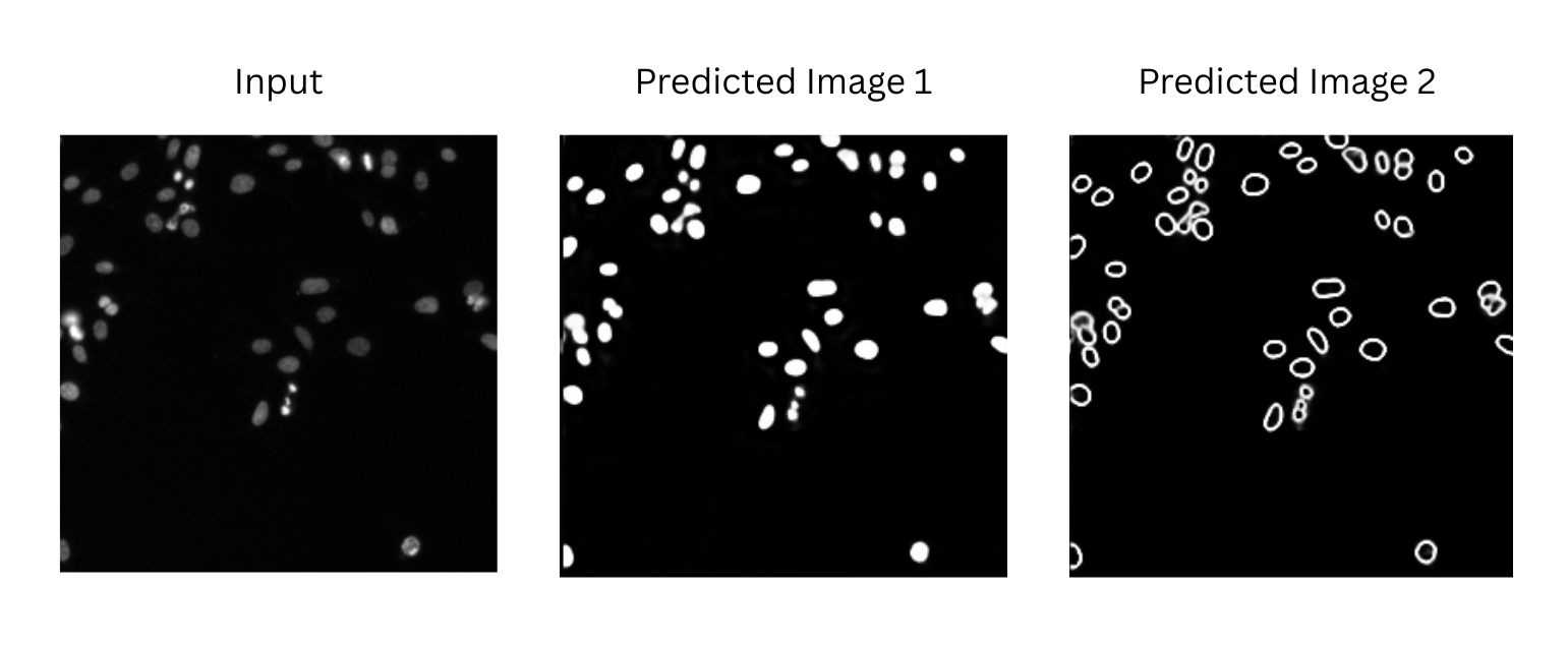
As an example, we consider the following model: 🧬 NucleiSegmentationBoundaryModel
This model segments nuclei in fluorescence microscopy images. It predicts boundary maps and foreground probabilities for nucleus segmentation, primarily in images stained with DAPI. The outputs are designed to be post-processed with methods such as Multicut or Watershed to achieve instance-level segmentation (object-based segmentation).
You can find similar details for other models directly on BioImage.IO by viewing each model’s card. Look under the “inputs” section of the RDF file to find the required axes and input size values. These parameters are essential for running the model correctly in Galaxy.
Get data
Hands On: Data Upload
Create a new history for this tutorial.
Download the following image and import it into your Galaxy history.
For the purpose of this tutorial, we will use one image to test only one of the 11 available models:
The input axes define the order of image dimensions expected by the model:
b: batch
c: channel
y: vertical axis
x: horizontal axis
The input size must match that order.
For example: 256,256,1,1 = 256 px height (y), 256 px width (x), 1 channel (c), and 1 image (b).
This information is provided in the model’s RDF file on BioImage.IO.
The model will process the input image and generate two outputs:
Two predicted images (written in one TIFF file)
A predicted tensor matrix (.npy)
Below is a visualization of the two predicted images generated by the 🧬 NucleiSegmentationBoundaryModel.
👉 See tip below for how to properly visualize the output.
Galaxy provides a basic preview using its .tiff visualization tool. However, BioImage.IO models sometimes produce tiff files with several predicted images residing in the same tiff file.
To properly explore the results, it is recommended to click on the visualize icon in the output file, this will give you the option to display the dataset using the Avivator tool.
Figure 2: Visualize your Tiff output with Avivator in Galaxy
An alternative is to download the file and open it locally using image analysis tools such as Fiji/ImageJ, napari, or QuPath.
Question: Check your understanding
Why do the image axes matter when using a model?
What happens if the image size does not match the model input?
What are TIFF and NPY formats?
How can you interpret the output of the model, and what does it tell you about your input image?
Because deep learning models are trained on specific image shapes and dimensions; mismatches will cause errors or wrong results.
The model will fail to run or produce invalid output.
TIFF (.tif) is a standard format for storing image data, commonly used in microscopy and bioimaging. It can be easily viewed and interpreted visually.
NPY (.npy) is a binary format used by NumPy to store arrays. In this case, it contains the raw prediction tensor produced by the model, which can be useful for further analysis or visualization with Python tools.
The model generates a predicted image that highlights or segments specific structures (e.g. nuclei, cells, mitochondria) based on what it learned during training. By comparing the output image to the input, users can see which regions were detected or classified, helping to extract biological meaning from the raw data.
Conclusion
In this tutorial, you learned how to run a BioImage.IO model on a biological image using Galaxy. By uploading a compatible model and image, setting the appropriate size and axes, and running the tool, you obtained both a predicted image and a tensor matrix representing the model output.
This provides a fast, reproducible way to apply deep learning models in the context of bioimage analysis — all within Galaxy.
You've Finished the Tutorial
Please also consider filling out the Feedback Form as well!
Key points
BioImage.IO models can be run in Galaxy using a dedicated tool
Input image and model need to be compatible in size and axes
Galaxy returns both the predicted image and its tensor as output
Frequently Asked Questions
Have questions about this tutorial? Have a look at the available FAQ pages and support channels
Further information, including links to documentation and original publications, regarding the tools, analysis techniques and the interpretation of results described in this tutorial can be found here.
References
Batut, B., S. Hiltemann, A. Bagnacani, D. Baker, V. Bhardwaj et al., 2018 Community-Driven Data Analysis Training for Biology. Cell Systems 6: 752–758.e1. 10.1016/j.cels.2018.05.012
Moen, E., D. Bannon, T. Kudo, W. Graf, M. W. Covert et al., 2019 Deep learning for cellular image analysis. Nature Methods 16: 1233–1246. 10.1038/s41592-019-0403-1
Gómez-de-Mariscal, E., C. García-López-de-Haro, W. Ouyang, L. Donati, E. Lundberg et al., 2021 DeepImageJ: A user-friendly plugin to run deep learning models in ImageJ. Nature Methods 18: 1192–1195. 10.1038/s41592-021-01262-9
Wei, D., X. Yi, S. Fong, A. M. Walczak, H. A. Demirci et al., 2021 BioImage.IO: A community-driven framework for AI model sharing in bioimaging. Nature Methods 18: 1196–1199. 10.1038/s41592-021-01333-x
Chamier, L. von, R. F. Laine, J. Jukkala, C. Spahn, D. Krentzel et al., 2021 ZeroCostDL4Mic: An open platform to simplify access and use of deep-learning in microscopy. Nature Communications 12: 2276. 10.1038/s41467-021-22518-0
Feedback
Did you use this material as an instructor? Feel free to give us feedback on how it went.
Did you use this material as a learner or student? Click the form below to leave feedback.
Hiltemann, Saskia, Rasche, Helena et al., 2023 Galaxy Training: A Powerful Framework for Teaching! PLOS Computational Biology 10.1371/journal.pcbi.1010752
Batut et al., 2018 Community-Driven Data Analysis Training for Biology Cell Systems 10.1016/j.cels.2018.05.012
@misc{imaging-process-image-bioimageio,
author = "Diana Chiang Jurado",
	title = "Using BioImage.IO models for image analysis in Galaxy (Galaxy Training Materials)",
	year = "",
	month = "",
	day = "",
	url = "\url{https://training.galaxyproject.org/training-material/topics/imaging/tutorials/process-image-bioimageio/tutorial.html}",
	note = "[Online; accessed TODAY]"
}
@article{Hiltemann_2023,
	doi = {10.1371/journal.pcbi.1010752},
	url = {https://doi.org/10.1371%2Fjournal.pcbi.1010752},
	year = 2023,
	month = {jan},
	publisher = {Public Library of Science ({PLoS})},
	volume = {19},
	number = {1},
	pages = {e1010752},
	author = {Saskia Hiltemann and Helena Rasche and Simon Gladman and Hans-Rudolf Hotz and Delphine Larivi{\`{e}}re and Daniel Blankenberg and Pratik D. Jagtap and Thomas Wollmann and Anthony Bretaudeau and Nadia Gou{\'{e}} and Timothy J. Griffin and Coline Royaux and Yvan Le Bras and Subina Mehta and Anna Syme and Frederik Coppens and Bert Droesbeke and Nicola Soranzo and Wendi Bacon and Fotis Psomopoulos and Crist{\'{o}}bal Gallardo-Alba and John Davis and Melanie Christine Föll and Matthias Fahrner and Maria A. Doyle and Beatriz Serrano-Solano and Anne Claire Fouilloux and Peter van Heusden and Wolfgang Maier and Dave Clements and Florian Heyl and Björn Grüning and B{\'{e}}r{\'{e}}nice Batut and},
	editor = {Francis Ouellette},
	title = {Galaxy Training: A powerful framework for teaching!},
	journal = {PLoS Comput Biol}
}
Congratulations on successfully completing this tutorial!
You can use Ephemeris's shed-tools install command to install the tools used in this tutorial.
Questions:
 Open image in new tab
Open image in new tab